

The Nobel Prize in Physiology or Medicine 2026 went to Karl Deisseroth, Peter Hegemann and Georg Nagel “for discoveries concerning light-gated ion channels and optogenetics.” The announcement was made on 5 October at the Karolinska Institute in Stockholm. Each laureate receives one-third of the prize. The total is 12 million Swedish kronor, roughly $1.2 million.
The award recognizes a technique that lets researchers switch specific nerve cells on or off with flashes of light, with millisecond precision. For most of the history of neuroscience, scientists could record from neurons or stimulate them with electrodes and drugs. They could not selectively control one defined cell type among billions of intermingled neighbours. Optogenetics solved that problem. It began with a curiosity about how single-celled green algae find sunlight...
Join Weekly Health Newsletter
Every week, I will share with you information about Natural Health, Diet, Exercise, Yoga & Wellness.
THE LAUREATES
Karl Deisseroth
Deisseroth is a bioengineer, neuroscientist and practicing psychiatrist at Stanford University. His laboratory turned microbial light-sensitive proteins into a general-purpose tool for neuroscience. He took the first step in 2005, in a collaboration that included Edward Boyden, Feng Zhang, Ernst Bamberg and Georg Nagel.
He then developed delivery methods, new opsin variants, fiber-optic light delivery for freely moving animals, and tools for imaging and mapping brain circuits. Because he treats patients with psychiatric illness, his work has been driven by the question of which circuits go wrong in disease and how they could be corrected.
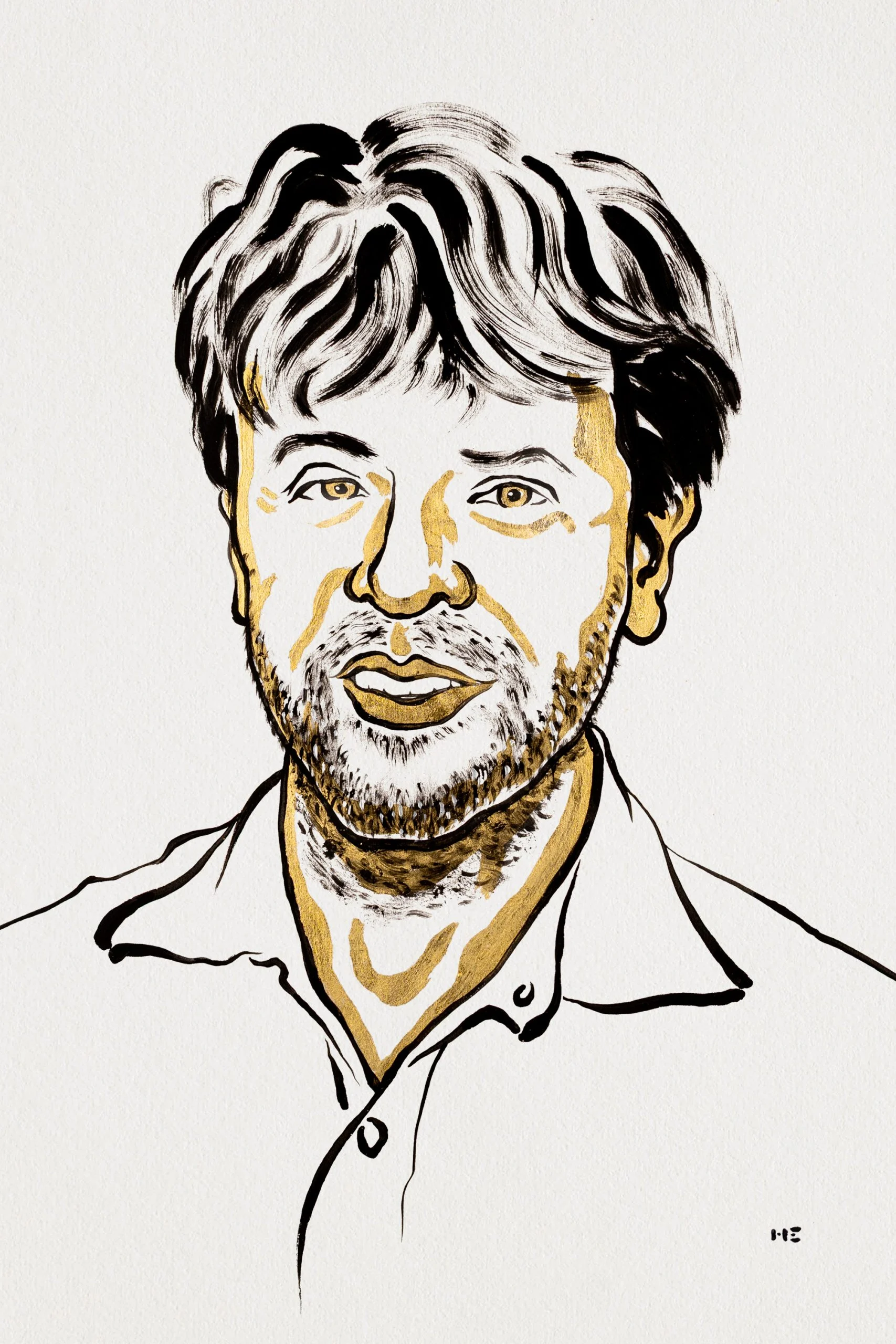

Peter Hegemann
Hegemann is a biophysicist at Humboldt University of Berlin. For decades he studied how the single-celled green alga Chlamydomonas reinhardtii senses light and swims toward or away from it.
This work, which seemed far removed from medicine, led him to propose that the alga’s photoreceptors might themselves be ion channels, which would make them directly gated by light.
He later expanded the family of known channelrhodopsins and engineered variants with new properties, including different colors of activating light and different ion selectivity.
Georg Nagel
Nagel is a biophysicist at the University of Würzburg. He worked on membrane transport and electrophysiology, and with Hegemann and Ernst Bamberg he showed that these algal photoreceptors are light-gated ion channels.
His electrophysiological skill allowed the team to demonstrate, in cells that normally do not respond to light, that illumination alone could open a channel and generate an electrical current.
A Note on Ernst Bamberg
Ernst Bamberg was a pioneer in membrane biophysics at the Max Planck Institute of Biophysics in Frankfurt and a central figure in the original discoveries.
He died in 2020. Nobel Prizes are not awarded posthumously, so his name is absent from the citation. His role is repeatedly acknowledged in the scientific literature.

THE SCIENTIFIC BACKGROUND
The Problem: Cell-Type-Specific Control of Neural Activity
The mammalian brain contains tens of billions of neurons from hundreds of molecularly distinct types, interleaved within the same tissue volume. Classical interventions lacked either specificity or speed. Electrical stimulation excites any element near the electrode, including axons of passage, and does not silence neurons selectively. Pharmacological agents are cell-type-selective only to the extent that their receptors are, and they act over seconds to minutes. Lesions are irreversible.
Francis Crick (1979) argued that neuroscience needed a method to control one cell type while leaving the others unperturbed, and he speculated that light might be a suitable control signal. Light offers temporal precision and spatial confinement, but neurons are not intrinsically light-sensitive. The solution required making specific neurons responsive to light through genetic means. Early attempts used multi-component systems, such as ectopic expression of the photoreceptor cascade or caged ligands (Zemelman et al., 2002; Lima & Miesenböck, 2005). These worked but were slow and technically demanding, and they depended on several exogenous genes.
Phototaxis in Chlamydomonas
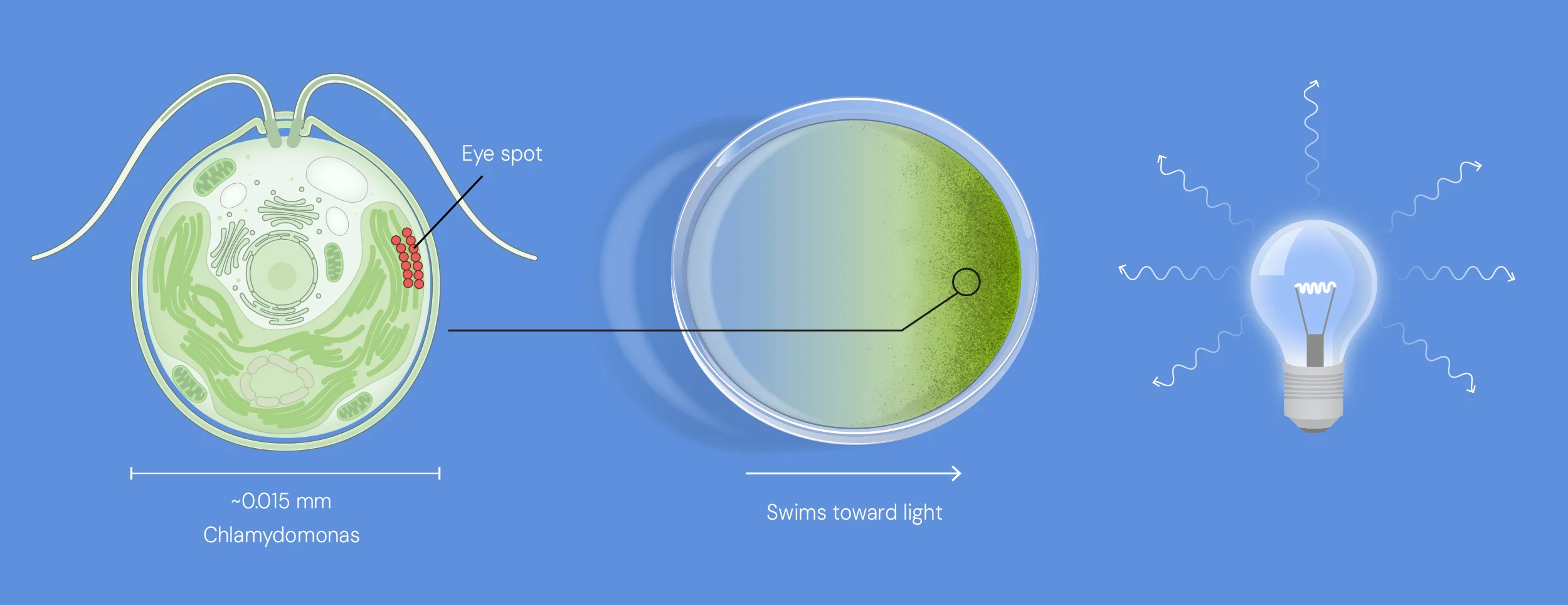
The alga Chlamydomonas senses light using its eyespot
The unicellular green alga Chlamydomonas reinhardtii swims toward or away from light using flagella. Foster and colleagues (1984) showed that phototaxis depends on a retinal-based photoreceptor, which implied a rhodopsin. Hegemann and colleagues then characterized the electrical events at the cell’s eyespot. Harz and Hegemann (1991) reported that light evokes photoreceptor currents with calcium-dependent components that precede the flagellar response by only milliseconds.
The latency was the key observation. A slow second-messenger cascade could not easily account for such rapid electrical responses. This raised the hypothesis that the photoreceptor might be directly coupled to an ion channel, or might itself be one. Sineshchekov, Jung and Spudich (2002) strengthened the case that rhodopsins mediated these currents, but the molecular identities and the gating mechanism remained unresolved.
THE DISCOVERY: CHANNELRHODOPSINS
Identification and Characterization of Channelrhodopsins
The genome and expressed-sequence-tag resources for Chlamydomonas revealed sequences with homology to the opsin domain of bacterio-opsin. Two were designated channelopsin-1 and channelopsin-2 (Chop1 and Chop2). The decisive experiments were heterologous expression studies, performed in the laboratories of Hegemann and Ernst Bamberg, with Georg Nagel contributing the electrophysiology.
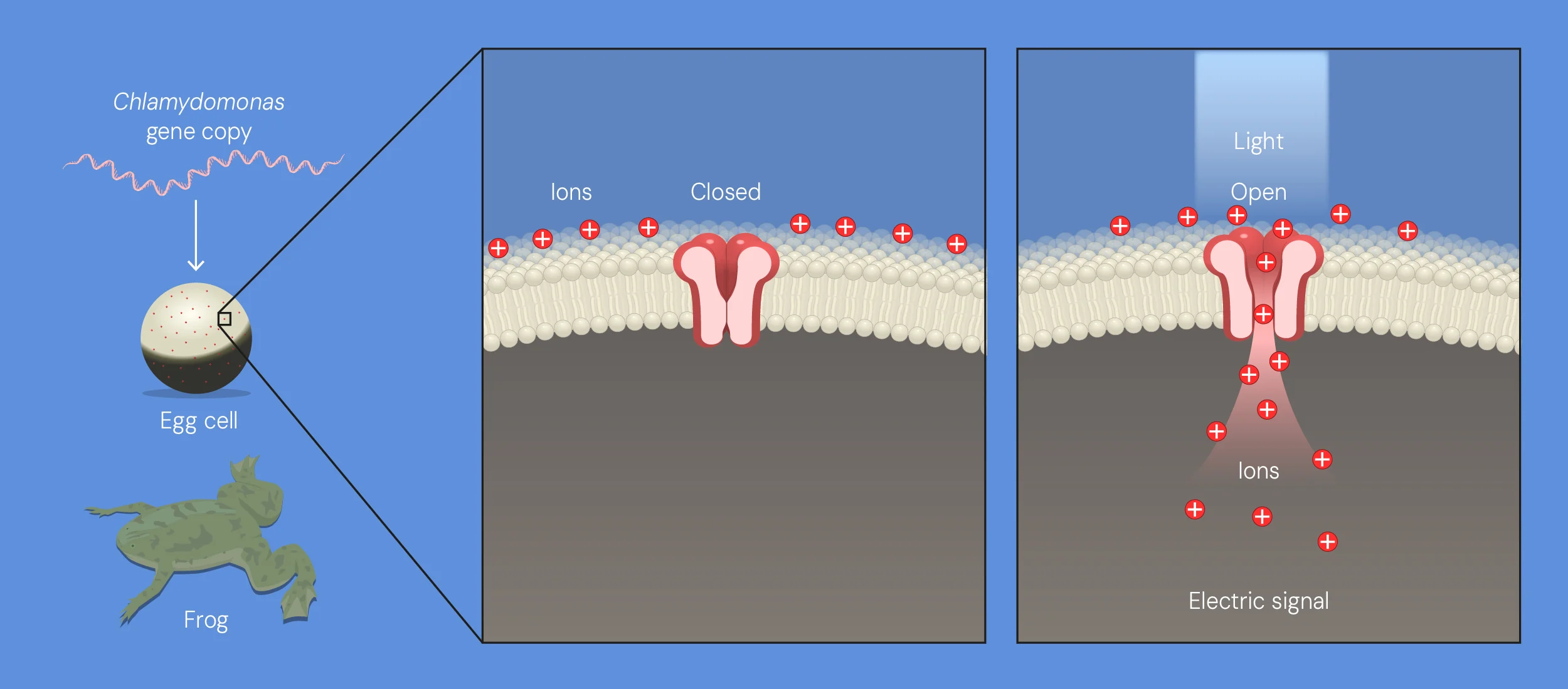
When expressed in Xenopus oocytes and mammalian (HEK293) cells, in the presence of all-trans retinal, these proteins produced light-activated membrane currents (Nagel et al., 2002, 2003). The key points were:
- Sufficiency. A cell with no algal machinery produced photocurrents. The N-terminal portion of the protein, containing the seven-helix rhodopsin domain, was sufficient. The photoreceptor and the channel are one polypeptide.
- Channelrhodopsin-1 (ChR1) conducted protons selectively (Nagel et al., 2002).
- Channelrhodopsin-2 (ChR2) conducted a mix of cations, including Na⁺, K⁺, Ca²⁺ and H⁺, with an absorption maximum near 470 nm (blue light) (Nagel et al., 2003). Its non-selective cation conductance depolarizes cells, which is the property that makes it useful for exciting neurons.
- Kinetics. Currents began within milliseconds of illumination, reached a peak, then declined to a smaller steady-state level (desensitization), and closed within milliseconds after light-off. The decay time sets the temporal fidelity with which the channel can follow pulsed light.
Structural work later supported a mechanism distinct from that of the pumps. The crystal structure of a ChR1–ChR2 chimera (C1C2) showed a hydrated, cation-conducting pore lined by several transmembrane helices (Kato et al., 2012). In the photocycle, isomerization of the retinal chromophore is followed by deprotonation and reprotonation of the Schiff base, spectroscopically distinct intermediates, and a conducting state that decays over milliseconds to tens of milliseconds. A caveat concerns throughput: unitary conductance of ChR2 is small, estimated in the tens of femtosiemens (Feldbauer et al., 2009). Producing large photocurrents therefore requires dense expression, and this has driven much of the later engineering.
Translation to Excitable Cells and Intact Organisms
ChR2 had several properties that made it a natural candidate for neuroscience. It is a single gene. It needs only retinal, which is present at sufficient levels in vertebrate tissue. It depolarizes cells. And its gating kinetics are fast.
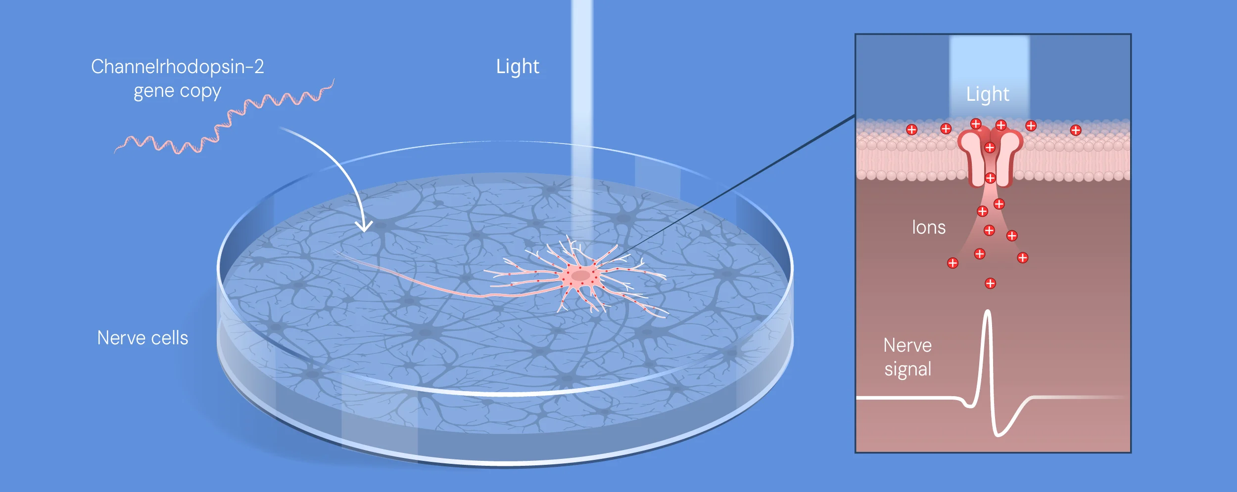
Boyden, Zhang, Bamberg, Nagel and Deisseroth (2005) expressed ChR2 in cultured hippocampal neurons using a lentiviral vector. Brief pulses of blue light evoked action potentials with millisecond-scale temporal precision, and trains of light pulses evoked spike trains at tens of hertz. The neurons’ normal properties were preserved, which indicated that expression was well tolerated.
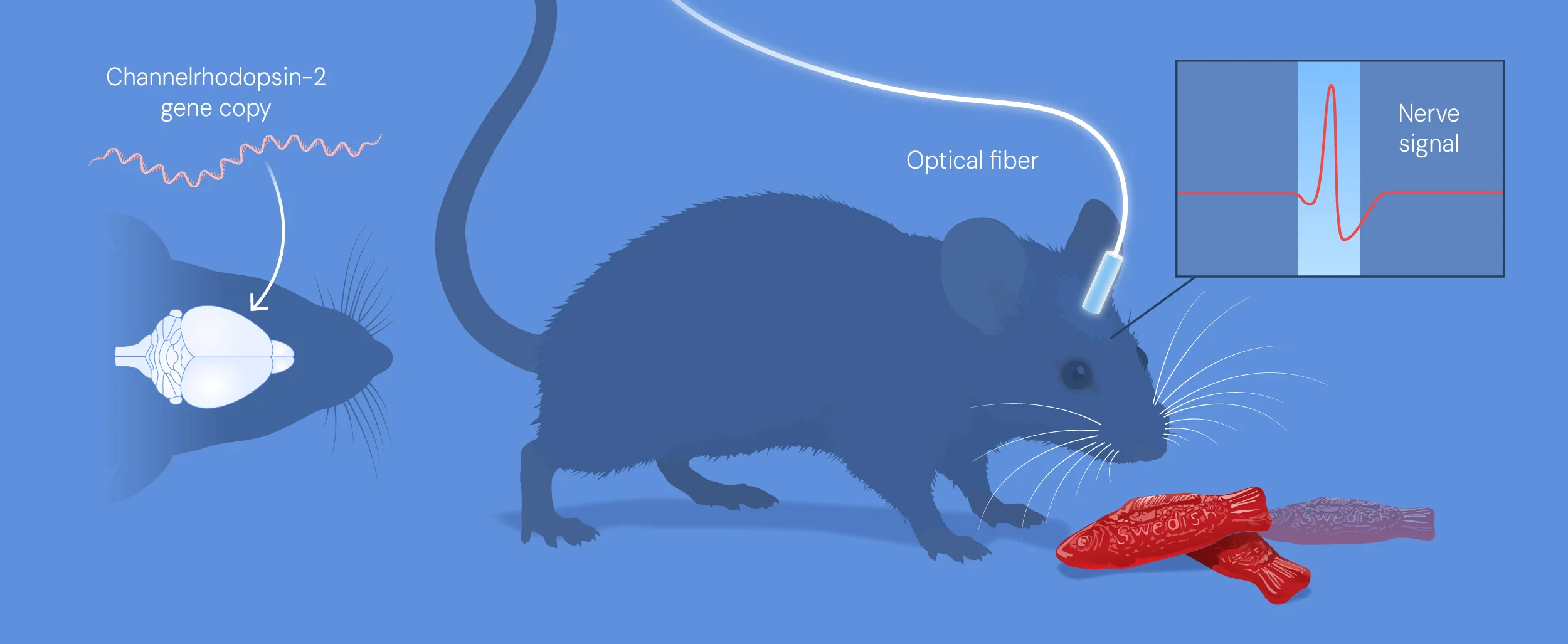
Independent work reported similar findings in parallel, including in neuronal networks (Li et al., 2005), in the nematode C. elegans, where light evoked behavior in intact animals (Nagel et al., 2005), and in retinal neurons (Bi et al., 2006). The convergence of these results established the approach.
OPTOGENETICS EXPERIMENT
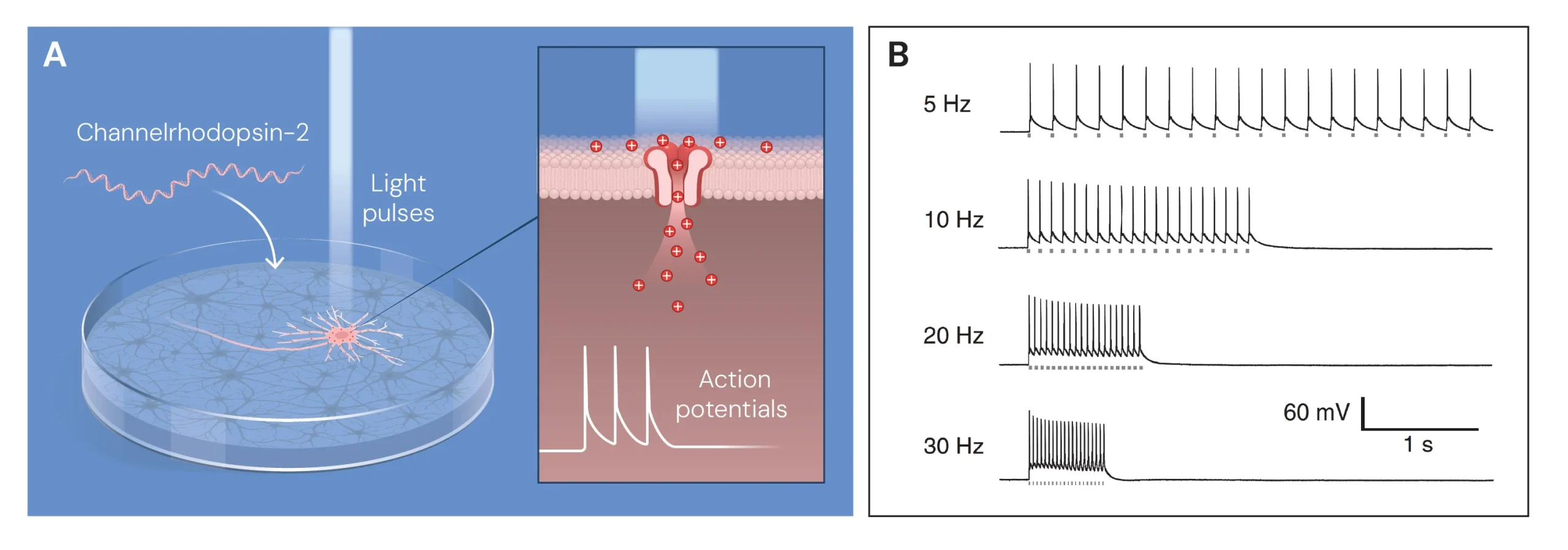
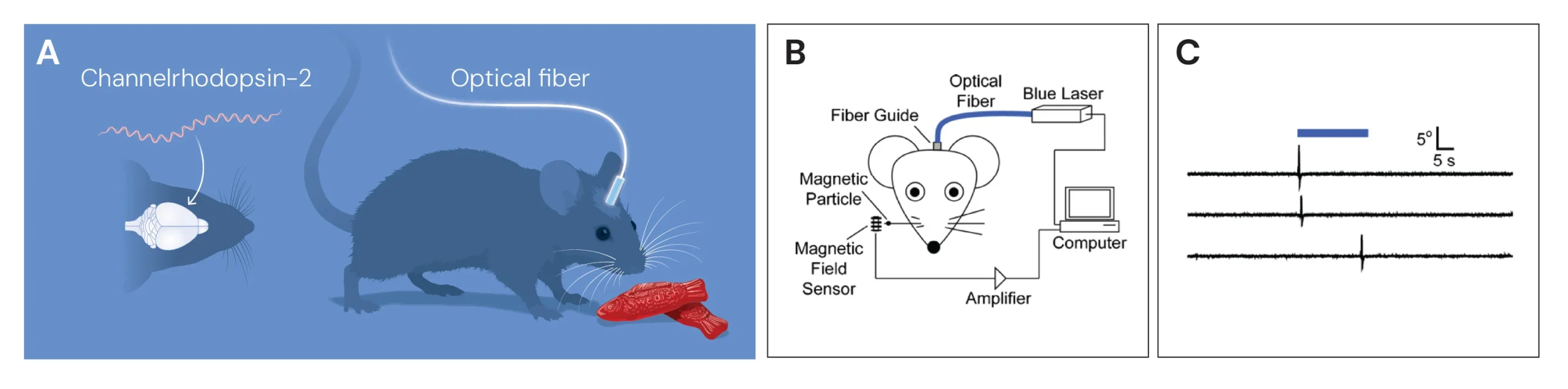
An optogenetic experiment combines four elements.
Actuators. The opsin is the effector, and the toolbox expanded rapidly after 2005 through discovery of natural variants and structure-guided engineering.
Genetic targeting. Specificity is achieved by driving opsin expression with cell-type-selective promoters, by using transgenic Cre-driver animal lines with Cre-dependent viral constructs, and by intersectional strategies that require two features at once (Fenno et al., 2014). Viral serotype and injection site add anatomical specificity, and projection-defined populations can be targeted by expressing the opsin in one region and illuminating the axon terminals in another.
Light delivery. In vivo work uses implanted optical fibers coupled to lasers or LEDs (Aravanis et al., 2007; Adamantidis et al., 2007). Typical irradiances are on the order of milliwatts per square millimeter. Because tissue scatters and absorbs blue light, the effective volume of tissue activated is limited, which motivated the red-shifted variants.
Readout. Optogenetic control is typically paired with electrophysiology, imaging or behavior. Combined with functional MRI, it allowed researchers to map brain-wide responses to activation of defined cells (Lee et al., 2010). Deisseroth’s laboratory also developed tissue-clearing methods that render intact brains transparent so that opsin-expressing circuits can be traced (Chung et al., 2013).
Tool Class | Example | Effect | References |
|---|---|---|---|
Blue-light cation channel | ChR2, ChETA | Depolarization, fast kinetics | Boyden et al., 2005; Gunaydin et al., 2010 |
Red-shifted cation channel | C1V1, Chrimson | Depolarization with deeper-penetrating light | Yizhar et al., 2011; Klapoetke et al., 2014 |
Bistable / step-function opsin | SFO | Prolonged activation after a brief pulse | Berndt et al., 2009 |
Light-driven pump | Halorhodopsin, archaerhodopsin | Hyperpolarization (silencing) | Zhang et al., 2007; Chow et al., 2010 |
Anion-conducting channel | Engineered ChloC, iC1C2; natural GtACRs | Inhibition by chloride flux | Wietek et al., 2014; Berndt et al., 2014; Govorunova et al., 2015 |
Light-activated GPCR chimeras | Opto-XRs | Control of intracellular signaling | Airan et al., 2009 |
Rigorous use of the method requires awareness of its artifacts:
- Non-physiological activation. Light synchronously activates all opsin-expressing cells, which can differ from natural activity patterns.
- Heating. Light absorption can raise tissue temperature enough to alter neural activity independently of opsin expression, which makes light-only controls essential (Owen et al., 2019).
- Inhibition is not always clean. Pump-based silencing can alter ion gradients and cause rebound effects, and silencing at presynaptic terminals has biophysical constraints that can produce unintended results (Mahn et al., 2016). This is one rationale for the development of anion-channel tools.
- Expression and specificity. Overexpression can affect cell health, and leaky recombinase expression can compromise targeting, so histological verification is necessary.
- Interpretation. A behavioral effect of stimulating a cell population shows that the population can drive the behavior. It does not by itself show that the population normally does so.
USES AND SIGNIFICANCE
Before optogenetics, much of systems neuroscience was correlational. Researchers could observe that a certain cell population became active during a behavior. They could rarely show it was necessary or sufficient for that behavior. Optogenetics made causal experiments routine. Researchers have used it to:
- Dissect circuits for fear, anxiety, reward, feeding and social behavior.
- Identify cell populations involved in sleep and arousal.
- Probe how memories are formed and retrieved by reactivating the neurons that encoded them.
- Study the circuits disrupted in movement disorders such as Parkinson’s disease, and clarify which pathways deep brain stimulation may be acting on.
- Map how seizures start and spread, and test ways to stop them.
Because opsins work in any excitable cell, the method has spread to other organs. Researchers have used light to control heart muscle, to study arrhythmias in experimental models, and to explore how sensory systems encode information. It has also become standard in studies of the spinal cord, the retina and the cochlea.
Understanding Psychiatric and Neurological Disease
Disorders such as depression, schizophrenia, addiction, epilepsy and Parkinson’s disease are increasingly understood as problems of circuits as much as chemicals. Optogenetics lets researchers test specific hypotheses about which circuits and cell types contribute to symptoms in animal models. This has informed the development of better-targeted treatments, including improved approaches to neuromodulation. It does not by itself cure these conditions, but it provides the causal evidence needed to focus therapeutic efforts.
Restoring Vision
The most advanced clinical application so far is in vision. In inherited retinal degeneration, light-sensing photoreceptors die while other retinal cells survive. Delivering an opsin gene to surviving cells can make them light-sensitive again. In 2021, a team reported that a patient with advanced retinitis pigmentosa regained partial, measurable visual function after receiving a gene therapy that encoded a channelrhodopsin variant, used together with specialized goggles. The result was modest and came from a single reported case, but it was an important proof of principle that optogenetic therapy can work in humans
Prospects and Hurdles
Other possible applications are under investigation, including optical control of hearing, targeted pain relief, and closed-loop systems that detect abnormal activity and respond with light. Several obstacles remain:
- Gene delivery: Viral vectors must reach the right cells safely, and the immune system may respond to them or to the foreign opsin.
- Light delivery: Light scatters in tissue, so deep brain applications require implanted devices.
- Safety and durability: Long-term expression, tissue heating and unwanted effects on cell health need careful study.
- Ethics: Any therapy that alters neural activity in the brain, even a well-intentioned one, will need rigorous oversight and informed consent.
0 comments