

Does the adult human brain keep making new neurons, and does that process falter in depression? Both questions have been argued over for decades. A 2026 Nature Medicine paper from Columbia University and the New York State Psychiatric Institute, led by Madeleine Peng, Jialin Jiang and Maura Dupont, tackles them with what the authors describe as the largest human hippocampus dataset to date.
Its central claim has two parts. The adult human hippocampus contains a recognizable neurogenic lineage, and in people with major depressive disorder (MDD) that lineage appears stalled rather than depleted. This article explains how the study worked, what it found, and what supports and limits it.
AT A GLANCE
Paper: Peng MS, Jiang J, Polizzi L, et al. Nature Medicine, 2026. doi:10.1038/s41591-026-04571-8
Scale: 495,037 nuclei from the dentate gyrus (DG) and cornu ammonis (CA) regions
Cohort: 123 postmortem donors (55 MDD, 68 controls), with different subsets used for each assay
Key condition: MDD cases were nonmedicated, with no psychotropic drugs in the three months before death except benzodiazepines
Core result: more early neural stem cells (NSCa) and fewer neuroblasts in MDD, with elevated interferon signaling in early neurogenic stages
Join Weekly Health Newsletter
Every week, I will share with you information about Natural Health, Diet, Exercise, Yoga & Wellness.
BACKGROUND
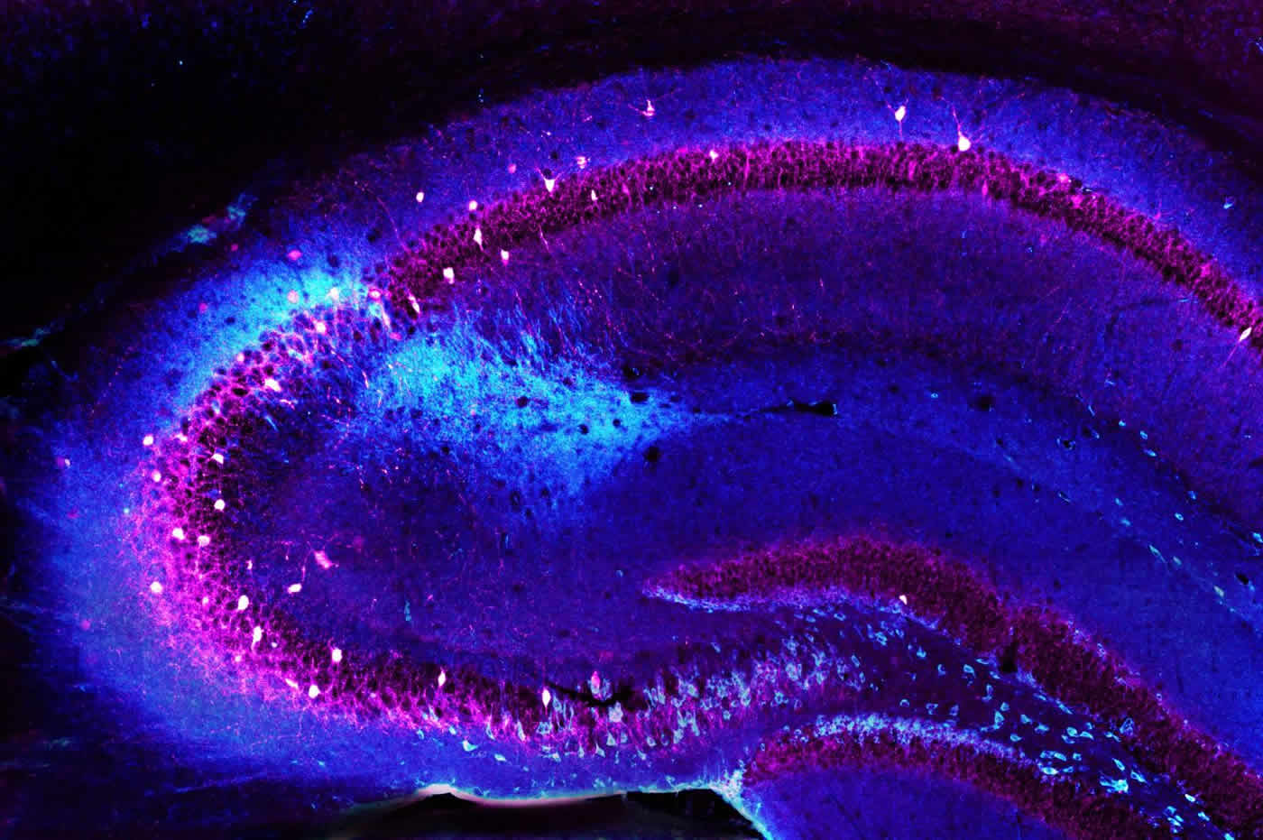
Why the Hippocampus?
MDD involves mood, neurovegetative and cognitive symptoms, including a bias toward recalling negative content. The paper notes that MDD is associated with reduced hippocampal volume, altered connectivity and negative memory bias, which suggests disrupted hippocampal plasticity.
The Neurogenesis Question
In rodents, adult hippocampal neurogenesis (AHN) supports pattern separation, the ability to tell similar experiences apart.
The authors cite a human study in which pattern separation became transiently deficient after focal hippocampal irradiation, which likely depletes AHN. Evidence that AHN occurs in the adult human brain has been growing, but the paper says its clinical relevance remains debated. It also says a molecular atlas of hippocampal circuit cell states in MDD was missing.
HOW THE STUDY WAS DONE
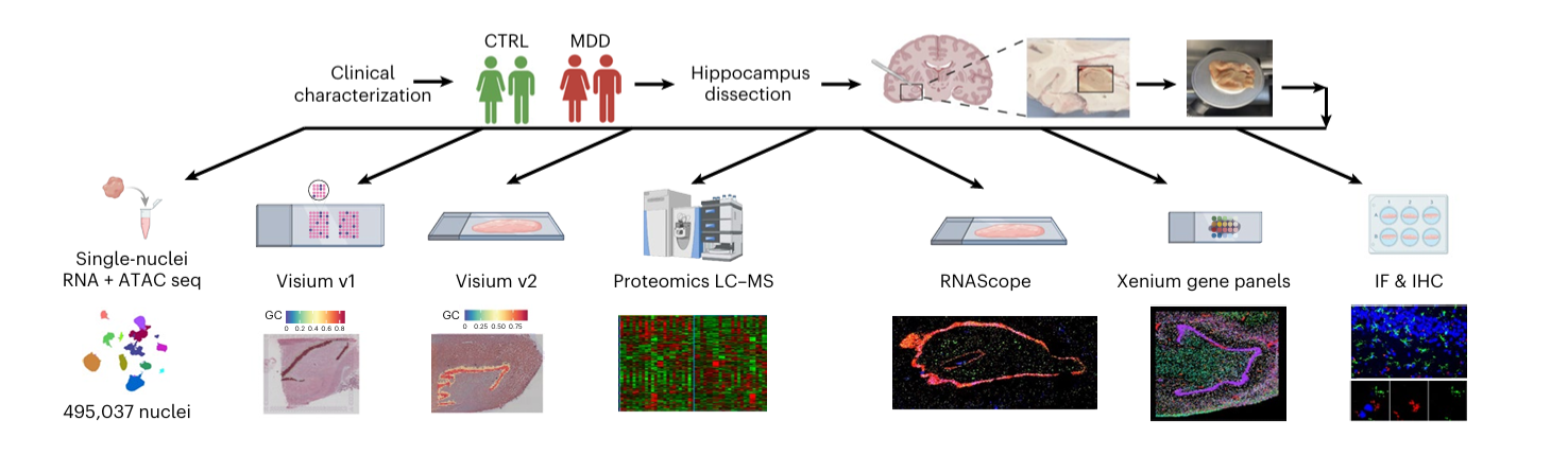
Schematic Study Design
The Cohort
Inclusion criteria were demanding: negative neuropathology, brain pH of at least 5.7, sudden death with a short agonal state, and negative toxicology for psychotropic drugs, alcohol and other substances. MDD cases met DSM criteria without psychosis, bipolar disorder or comorbid substance or alcohol use disorders. Controls had no psychiatric diagnosis, treatment or history of suicide attempts.
The sample sizes differ by assay:
- Single-nucleus multiome (snMultiome): 11 MDD and 19 control donors after quality control, giving 74 samples with 1–4 technical replicates per donor
- Visium spatial transcriptomics: 13 control and 14 MDD donors, according to the Methods
- Proteomics: 12 MDD and 12 controls
- Validation: Xenium, RNAscope, immunohistochemistry and immunofluorescence on larger subsets, for example nestin in 71 samples and doublecortin in 58
Overall, 72.7% of the MDD group died by suicide (40 of 55) versus none of the controls. About 72% of donors were male. MDD cases had higher stress exposure in the last six months of life on the St. Paul–Ramsey scale (2.4 versus 1.8).
The Technologies
Tissue came from the anterior hippocampus proper, including DG and CA regions. The team combined:
- snMultiome-seq: gene expression and chromatin accessibility in the same nuclei
- Spatial transcriptomics (Visium v1/v2 and single-cell Xenium): to place cell types and trajectories anatomically
- Regional proteomics (LC-MS/MS): to check changes at the protein level
- Computational inference: trajectory tools (Monocle3, Palantir, RNA velocity), transcription factor footprinting (TOBIAS) and regulon analysis (pySCENIC)
- Tissue validation: immunohistochemistry, RNAscope and immunofluorescence with stereological cell counts
KEY FINDINGS
A Large Atlas of the Adult Human Hippocampus
The team identified 31 cell clusters, including granule cells (GCs), GABAergic and excitatory neurons, and astrocytes, and mapped them onto hippocampal subfields. One astrocyte cluster (Astro2) was enriched in the neurogenic subgranular zone (SGZ). The authors also describe a previously uncharacterized PENK+ medium-spiny-neuron-like GABAergic cluster (InN5.PENK).
Existence of a Neurogenic Lineage in Adults
By sub-clustering astrocyte and GC populations, the authors found a continuous lineage: quiescent neural stem cells (NSCa), activated stem cells (NSCb), intermediate progenitors (INPs), neuroblasts (NBs) and two immature granule cell types (ImGC1, ImGC2). Three further lines of evidence support it:
- Fetal similarity: adult neurogenic clusters co-embedded with fetal hippocampal progenitors
- Spatial trajectory: RNA velocity on Visium data suggested movement from the SGZ toward the granule cell layer
- In situ markers: Xenium and immunostaining detected stem-cell and neuroblast markers in the SGZ
The trajectory branches after ImGC1, with one route to mature GCs and another to ImGC2. ImGC2 cells sit in the granule cell layer rather than the SGZ and express immaturity genes such as BHLHE22, POSTN and FST. The authors read a possible path from mature GCs back to ImGC2 as “dematuration,” a phenomenon previously described in marmosets.
Stage-Specific Transcription Factors Steering the Lineage
Footprinting showed stemness regulators (SOX2, PAX6, TLX) in stem cells, ASCL1 and Notch-related factors (HES1, HES5, HEY1) in activated stem cells, and homeobox families in INPs. In GCs, binding converged on KLF15, a glucocorticoid-regulated factor. BHLHE22, whose gene has a variant linked to severe depression in veterans, was most active at the ImGC2 stage.
Neurogenesis in MDD
Pseudotime analysis showed more NSCa and fewer NBs in MDD, and stage-associated gene signatures were delayed in stem cells and lower in neuroblasts. The authors interpret this as impaired progression rather than stem-cell depletion.
The tissue validation backs this up:
- Fewer Ki67+ and nestin+ cells in the anterior DG of MDD cases
- Fewer doublecortin-expressing cells in the anterior DG
- No difference in ImGC2 marker cells between groups
The effects were concentrated in the anterior hippocampus. Mid and posterior DG counts were mostly similar between groups.
Interferon Signalling as a Candidate Brake
In early stages (NSCa, NSCb, INP), MDD cells showed higher expression of a gene module enriched for interferon α/β signaling and antiviral pathways. It included EIF2AK2, IFIT3, HERC6 and NRIR. Later stages showed other changes:
- Lower NELL1, POSTN and ADAMTS genes in INPs
- Higher SOX9, which favors a glial fate, in INPs
- Lower DCX and BDNF in neuroblasts
Immediate-early genes (FOS, JUN, EGR1) and heat-shock proteins were up in neurogenic cells, consistent with an enhanced stress response.
The Whole Circuit is Affected
Dysregulation was not confined to neurogenic cells. InN5.PENK had the most differentially expressed genes (566), followed by GC1 granule cells (378) and an excitatory CA cluster, ExN1.CA1-4.FIBCD1 (147). Shared themes included:
- Serotonin receptor genes (lower HTR4 and HTR7 in progenitor stages, lower HTR2A and HTR2C in spatial subfields, higher HTR1D in InN5.PENK)
- Downregulated ionotropic and metabotropic glutamate receptors
- Disrupted intracellular trafficking, apolipoprotein genes and long noncoding RNAs
- Interferon signaling also up in mature GCs and excitatory neurons
InN5.PENK also overexpressed proenkephalin and activity-related genes. The authors suggest this population might be compensating for a stress-hyperactivated DG.
Proteomics
Proteomics identified 297 differentially expressed proteins, including reduced calbindin. Several genes were dysregulated at both RNA and protein level, such as RAB6B, CLTCL1, NPTN, CAMKV, CYFIP2 and HPCAL1, pointing to trafficking, development and neurotransmission. HOMER1 was an interesting discordance: lower RNA but higher protein, which the authors speculate may be compensatory.
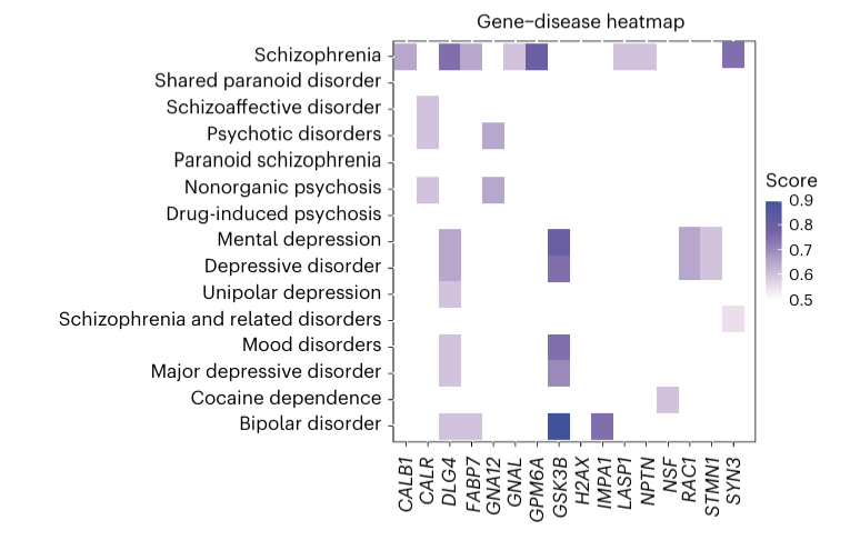
Gene-Disease Heatmap
Epigenetic and Stress-Related Regulation
Chromatin analysis found 102 differentially accessible regions in MDD, some overlapping differentially expressed genes. Footprinting highlighted KLF15 and other stress-responsive Krüppel-like factors as drivers of expression changes in GC1, InN5.PENK and ExN1. KLF15 binding correlated with expression of targets including HPCAL1, KRAS, CAMKV and RAB6B. This fits the observation that MDD donors had higher recent stress exposure, though the paper does not claim to prove causation.
Overlap with Other Conditions
Some differentially expressed genes are GWAS hits for depression (SAMD5, CSMD3, CDH9, TRHDE). Others have links to neurodevelopmental, neurodegenerative, autoimmune and thyroid-related conditions. Proteins such as GSK3β, STMN1, NPTN, CALB1 and FABP7 have also been implicated in schizophrenia, bipolar disorder and addiction. The authors read this as shared plasticity and development pathways across psychiatric disorders.
WHAT'S THE SIGNIFICANCE?
A cellular mechanism candidate. Reduced hippocampal volume is an old finding, but volume is a blunt measure. This work points to specific cell states and regulators.
A bridge to cognition. If stalled neurogenesis impairs pattern separation, it could help explain negative memory bias. The authors frame this as a new understanding of hippocampus-dependent cognitive symptoms, not a demonstrated mechanism.
Toward precision psychiatry. The paper says MDD pathways may “work in concert or represent different pathogenetic mechanisms of MDD subtypes.” It positions the work as a base for biologically informed diagnostic subtyping, biomarker discovery and new therapeutic targets.
A shared resource. Processed data are deposited on Zenodo and code is on GitHub, and the paper is open access.
STRENGTHS AND WEAKNESSES
What Supports the Paper
- Convergent methods. Gene expression, chromatin, spatial data, proteomics and tissue staining all point the same way for the lineage and the stall.
- Scale and depth. Extended Data Table 2 compares 495,037 nuclei and deep sequencing against earlier human hippocampus studies, which range from roughly 22,000 to 403,000 nuclei. Only one earlier study also included ATAC sequencing.
- Spatial and fetal anchoring. Placing neurogenic cells in the SGZ, showing a trajectory toward the granule cell layer, and matching adult cells to fetal progenitors address the common criticism that dissociated nuclei lose context.
- Cleaner MDD group. Excluding recent psychotropic medication, alcohol and substance use disorders, and psychosis or bipolar cases, removes major confounders.
- Independent validation. Protein and RNA staining in separate donor sets, analyzed by stereology, did not rely on the sequencing alone.
- Fit with the wider literature. The paper builds on recent work identifying proliferating neural progenitors and neurogenesis in adult human hippocampus (Dumitru et al., Science 2025; Disouky et al., Nature 2026).
- Transparency. Data, code and reporting summary are available, and competing interests are minimal.
What are the Limitations
As stated by the author:
- Cross-sectional, postmortem design. A snapshot after death cannot show onset, sequence or causality.
- Suicide confound. Most MDD donors died by suicide, so the authors acknowledge limited ability to separate MDD from suicide pathology.
- Modest power for adversity effects. Stress and adversity-related expression changes could not be examined well.
- Cause of death and unrecognized illness. These may have influenced the results.
As per my opinion:
- Association, not mechanism. No experiment tests whether interferon signaling or KLF15 actually causes the stall, or whether reversing it helps.
- Small primary sample for a heterogeneous disorder. Eleven MDD and nineteen control donors in the main single-nucleus analysis limits subtype or sex-specific conclusions. Technical replicates were handled statistically, but they do not add independent donors.
- Sex imbalance. About 72% of donors were male and only a third of the MDD group was female. Sex was a covariate, but findings may not generalize equally.
- Rare cells and lenient thresholds. For NSCs, INPs and NBs, the authors used unadjusted P < 0.05 with log₂FC > 1 “to reduce type II error due to low cell numbers.” This is understandable but raises false-positive risk. The interferon finding therefore deserves independent replication.
- Region and age range. Only the anterior hippocampus of the right hemisphere was studied, and MDD donors were 28–66 years old. Older adults and other regions are not covered.
- Literature-based interpretation. Many gene functions are inferred from cancer, autism, lupus and other contexts, which makes some mechanistic readings speculative.
- Retrospective stress measure. Stress exposure was reconstructed after death, and the paper itself does not tie it causally to the molecular changes.
WHAT'S NEXT?
Reasonable next steps include replicating the findings in independent brain banks with more donors and more non-suicide MDD cases. Functional tests of candidate drivers in human stem-cell-derived models or animal systems would address causality. Linking cognitive measures such as pattern separation to molecular signatures in living patients would test the memory hypothesis. Finally, researchers can ask whether a “stalled neurogenesis” signature defines a distinct biological subtype of depression.
0 comments