

If you learned embryology the way most of us did, the story of the brain starts simply. An early sheet of tissue becomes the nervous system, and that single sheet is gradually carved into forebrain, midbrain and hindbrain. A recent Nature Neuroscience paper from Stanford Medicine, led by Kyle Loh with Carolyn Dundes and Rayyan Jokhai as co-first authors, challenges the "single sheet" part of that story. Its title is "Two parallel neural ectoderm progenitors contribute to the developing brain," and the claim is that two distinct progenitor populations appear side by side during gastrulation, one fated for the front of the brain and one for the back.
This matters in developmental neuroscience for two reasons. First, it changes how we think about when brain regions become distinct, which affects how we interpret congenital malformations and regional vulnerability. Second, it has immediate practical consequences for stem cell biology: if the brain really has two separate starting points, protocols designed around one of them may never have been well suited to making the other.
Headlines have called this "two brains" or "two separate organs." That framing is catchy but overstated, and part of my goal here is to separate what the data show from what the headlines imply. Throughout, I will flag what is established, what is emerging, and what still needs to be checked against the primary paper.
Join Weekly Health Newsletter
Every week, I will share with you information about Natural Health, Diet, Exercise, Yoga & Wellness.

From Ectoderm to Brain
By roughly embryonic day 7.5 (E7.5) in the mouse, the ectoderm has sorted into three broad territories: neural ectoderm, which will form the brain; border ectoderm, which contributes neural crest; and surface ectoderm, which becomes the epidermis. This is well established and appears in standard developmental biology references.
The neural territory is marked by expression of the transcription factor Sox2. Later in development, cells adopt more specific neural progenitor identities and express markers such as Sox1 and Pax6. The new paper concerns the stage before that, when the neural ectoderm is still early and has not yet turned on those later markers.
Eventually, the neural tissue folds into the neural tube, which patterns into the forebrain (prosencephalon), midbrain (mesencephalon) and hindbrain (rhombencephalon). The hindbrain is further segmented into units called rhombomeres. These structures are the anatomical backdrop for everything below.
The Traditional Model: One Sheet, Divided Later
The conventional view, as described in coverage of the paper, is that the neural ectoderm first forms as a single continuous sheet and is only later subdivided into anterior (front) and posterior (back) regions. In this picture, a common brain progenitor can in principle generate all brain regions, and regional identity is imposed afterwards by signaling gradients.
This model has deep roots. Classical embryology experiments and later models of neural patterning described how signals cause neural tissue to adopt anterior or posterior character. It also shaped laboratory practice. Many stem cell protocols assumed that if you could coax pluripotent stem cells into a generic neural progenitor, you could then tune the recipe to produce whichever brain region you wanted.
That assumption worked reasonably well for some regions. Researchers have had considerable success converting human pluripotent stem cells (hPSCs) into forebrain and midbrain cells. The hindbrain, though, has been harder to produce, and the new work offers a possible explanation.
What "Two Parallel" Progenitors Actually Means
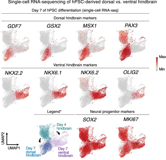
The central idea is this: rather than one progenitor population that later splits, two populations emerge at the same time during gastrulation and remain separate.
The authors call them:
- Anterior neural ectoderm (aNE): marked by Sox2 together with Otx2, and associated with the forebrain and midbrain.
- Posterior neural ectoderm (pNE): marked by Sox2 together with Gbx2, and associated with most of the hindbrain. The posterior population also expressed additional posterior markers, including Hoxa1 and Hoxb1.
In E7.5 mouse embryos, these two markers, Otx2 and Gbx2, were reported to be mutually exclusive within the Sox2-positive neural ectoderm.
In other words, a given early neural cell was either anterior-like or posterior-like, not a blend.
The word "parallel" is doing real work here. It contrasts with a "branching" model, in which posterior progenitors would split off later from anterior ones. As summarized in secondary coverage, the evidence presented points to the two arising separately from an early stage rather than one descending from the other.
It is also worth noting what these populations are not. The authors define aNE and pNE as neural ectoderm that arises during gastrulation, and they distinguish it from the forebrain, midbrain and hindbrain neural progenitors that emerge after gastrulation. So this is a claim about a very early developmental stage, before the familiar regional progenitors exist.
The authors describe the brain as potentially a "composite organ" emerging from two lineage-restricted progenitors. That phrase is the paper's careful wording. It is a long way from saying there are two independent brains.
How the Researchers Tested the Idea in Mouse Embryos
To ask where cells end up, you need lineage tracing, a technique that permanently labels cells at a given time so their descendants can be identified later. The study used lineage tracing in mouse embryos, including a Gbx2-driven inducible Cre system paired with a fluorescent reporter (described in secondary coverage as Gbx2-CreER crossed with a tdTomato reporter).
The headline numbers, as reported in summaries of the paper, are:
- 494 neural ectoderm-derived cell clusters were tracked across 16 independent embryos.
- About 63% of clusters (62.96%) were found exclusively in the forebrain and midbrain.
- About 33% (32.59%) were found exclusively in the hindbrain.
The key conceptual result is that descendants of the anterior and posterior populations mostly stayed in their respective territories. That supports the idea of two lineages with distinct developmental destinies, though, as with all lineage tracing, the conclusions depend on the specificity of the genetic labeling tools.
Testing Lineage Commitment With Human Stem Cells
Mouse embryos tell us about in vivo fate. To test whether the two populations are truly committed, meaning unable to switch identity, the researchers turned to human pluripotent stem cells.
They differentiated hPSCs into anterior or posterior neural ectoderm-like cells and found that these were lineage committed to forebrain/midbrain versus hindbrain fates, respectively. The more striking experiment was a kind of challenge test. When the cells were exposed to signals that would normally push them toward the opposite fate, they did not comply. Hindbrain-fated progenitors given forebrain-inducing signals activated hindbrain regulatory programs instead, and the reverse was reported for forebrain-fated progenitors.
The authors suggest these populations cannot transdifferentiate, meaning they cannot convert directly from one fate to the other. Importantly, this comes from an in vitro system. It tells us about the properties of these stem cell-derived populations, and it is a model of human development rather than direct observation of it. Early human embryos are not accessible for this kind of experiment, so stem cell models are the practical alternative, but they come with the usual caveat that a dish is not an embryo.
what are the implications?
Chromatin: A Possible Mechanism for Early Commitment
What might hold these fates in place? The study reports that anterior and posterior neural ectoderm have divergent chromatin landscapes, assessed with chromatin accessibility profiling (the datasets deposited for the study include ATAC-seq and single-cell RNA-seq). The authors describe these landscapes as foreshadowing the forebrain versus hindbrain potentials of the two populations.
In plain terms, even before overt regional structures appear, the cells' DNA is already organized differently, making some regulatory programs available and others harder to reach. This offers an attractive explanation for why signals that normally redirect cells failed to do so.
Why the Hindbrain Matters: A Practical Payoff
The hindbrain houses structures involved in functions essential to life, and the study's motivation was to generate human hindbrain cells from stem cells. Using their posterior neural ectoderm populations, the researchers differentiated hPSCs into hindbrain rhombomere 5/6-specific motor neurons.
Loh summarized the significance in a statement quoted by Epocrates: neurons from the back of the brain can now be grown in a dish and studied. The reasoning follows from the model. If earlier protocols were effectively aimed at the anterior progenitor, they would be poorly suited for producing posterior fates, and starting from the right progenitor opens a more direct route.
An Evolutionary Twist
The authors also raise an evolutionary hypothesis. They suggest that anterior and posterior neural ectoderm may be conserved from vertebrates to hemichordates, implying an ancient neural lineage bifurcation that predated chordates. The paper puts the possible age of this arrangement at roughly 550 million years.
Loh has framed this in interviews as evolution taking two existing neural systems and bringing them together spatially into one organ. It is an intriguing idea, and it fits with the observation that the anterior-posterior axis is a deeply conserved feature of animal body plans.
Still, this part of the work is a hypothesis built on comparative evidence. A summary of the study in a science-news repost is explicit about this: the strongest evidence concerns two distinct developmental programs, while the deeper evolutionary interpretation remains speculative.
conclusion
The central idea of this research is that the brain may not descend from one common neural progenitor that is later subdivided. Instead, two parallel neural ectoderm progenitors, one anterior and one posterior, appear together during gastrulation, carry distinct chromatin landscapes, and appear lineage-committed toward forebrain/midbrain versus hindbrain fates. If the model holds up, it reshapes how we think about early brain patterning, explains why stem cell recipes for one region did not transfer to another, and offers a more direct path to making human hindbrain neurons for study.
At the same time, the finding is best understood as an emerging model rather than settled textbook knowledge. The strongest data come from mouse lineage tracing and stem cell experiments, the evolutionary interpretation remains a hypothesis, and clinical relevance is still to be demonstrated.
0 comments